phytopthora POMEWEST
ON FARM PHYTOPHTHORA
BAITING
for early disease detection
Phytophthora species are serious plant pathogens, causing significant damage to commercial crops and natural systems across Australia.
Words Jennifer Riseley, Project Officer, Pomewest
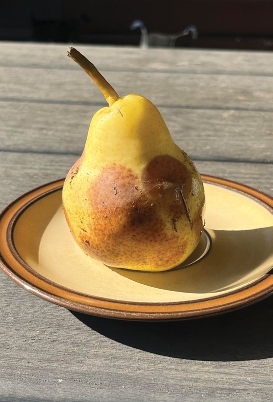
A pear showing lesions from both Phytophthora on the left side and Pythium on the right.
THERE are more than 60 species or taxa present in Australia (Burgess et al., 2017), many of which impact biodiversity, agriculture and horticulture. Though often called fungi, Phytophthora are funguslike organisms that share some characteristics with fungi. Early detection of Phytophthora in apple and pear orchards, even before symptoms are visible in trees, is essential to minimise production losses.
Phytophthora green pear baits can be used as an inexpensive, early detection method that isolates Phytophthora species from soil.
If the pathogen is present in high amounts, then symptoms on the pear baits will generally appear within 3–8 days. The method is also useful for distinguishing Phytophthora from other pathogens such as Pythium or true fungi. The test involves placing a pear in a flooded soil sample and waiting for the pathogen to infect the fruit skin.
Phytophthora zoospores can swim through water to find a host.
On farm testing guide
Here, we outline a user-friendly guide for growers to undertake this testing themselves.
Sample soil and roots soon after irrigation or rain so that soil is moist. Processing the root or soil sample should happen as soon as possible to stop the sample drying out and being attacked by other organisms. Avoid refrigerating the sample as this will reduce the recovery of Phytophthora and may give a false negative result.

FIGURE 1: Bath ring symptoms after 7 days of incubation – low level of inoculum in soil
Source: Tonya Wiechel, Agriculture Victoria
Materials required
- Heavy duty ziplock bags or clean plastic containers, wash and rinse if new, wash rinse and sanitise if reusing
- Pears, clean and without wounds or scald
- Unchlorinated water, bottled water suits well
- Space for samples to incubate at room temperature
Method
- Place soil sample in ziplock bag or plastic container. Multiple bags may be required in case of leakage.
- Make a well in the soil sample, either with clean gloves or through the plastic bag. Do not touch directly with hands and take care to avoid cross contamination.
- Carefully place the pear in the soil sample so that it stays in place, either upright or at 45 degrees, to ensure good water contact. Take care to avoid wounding the pear skin if the sample has any jagged material or rocks.
- Add enough water to flood the sample so the water line is on the neck of the pear. Water should be at room temperature (18–25ºC) or cooler and have no chlorine.
- Keep the water line topped up until symptoms appear, if using the original zip lock bag, keep the top open.
- Zoospores may be released in the first hour, or may take several days to mature. Leave the pear in the flooded sample for 20 days. Inspect daily to be sure bags are not leaking and check for symptom development. As soon as distinct lesions are visible at the water line (see Figure 1), remove the pear from the sample to avoid secondary infection from other pathogens.
Infections can occur anywhere on the submerged portion of the fruit (see Figure 2).
- Low levels of inoculum, may see isolated lesions at water line or where soil is in contact
- Medium inoculum, may see a ‘bathtub ring’ at the water line
- High levels, may see brown lesions covering base of fruit within 3 days

FIGURE 2: (a) Pear baiting set up day 0,

(b) after 7 days
Source: Tonya Wiechel, Agriculture Victoria
Identifying lesions
Generally, Phytophthora lesions are limited to the surface of the skin and remain firm to the touch. While submerged, the lesions may only be subtle in colour until removed from water where they oxidise and darken within the hour.
The lesions of Pythium and true fungi can sometimes be mistaken for Phytophthora.
Differences between the three lesions can be distinguished, as follows.
Phytophthora (see Figure 3):
• Firm dark lesions
• Lesions occur in non-wounded tissue, often a bathtub ring at water level
Pythium (see Figure 4):
• Often infect at a wound
• Lesions are soft and expand over time
• Watersoaked or translucent when in water
• Can expand quickly or slowly
• Can be dark brown, when surface of skin peeled back a soft decay of next level is seen
Fungi:
• Lesions typically at wounds
• Become sunken quickly
• Opaque lesions, not translucent
• Dry and crack after a few days

FIGURE 3: Lesions typical of Phytophthora
Source: Phytosphere.com

FIGURE 4: Lesions typical of Pythium
Source: Phytosphere.com
MORE INFORMATION
The Integrated Pest Disease and Weed Management manual for Australian Apple and Pears provides further information on the three species of Phytophthora most commonly associated with disease in Australian orchards. The manual provides detail on the disease, its impact in orchards, and how to prevent or manage infection. The manual can be downloaded free of charge from the Australian Apple and Pear IPDM web-site on extensionAus.

Or contact Dr Tonya Wiechel at Tonya.Wiechel@agriculture.vic.gov.au.
Acknowledgement
The PIPS 4 Profit program’s Integrated Pest and Disease Management project (AP22001) has been funded by Hort Innovation, using the apple and pear research and development levy, contributions from the Australian Government, and co-investment from Agriculture Victoria, and Pomewest in WA.
With thanks to Dr Tonya Wiechel of Agriculture Victoria.
References
Burgess, T., et al., (2017). Distribution and diversity of Phytophthora across Australia. Pacific Conservation Biology, 23, 1–13. https://doi.org/10.1071/PC16032
Phytosphere.com